An Introduction to Tumor Grading and Cancer Staging
Cancer is one of the most significant diseases worldwide. In 2018, over 1.7 million people were diagnosed with cancer in the United states. Moreover, approximately 38.4% of men and women are expected to be diagnosed with cancer in their lifetime (National Cancer institute). A critical component for cancer treatment is proper diagnosis of the tumor and severity of the disease. This can be a difficult process given the wide range of cancer types, tumor classifications and individual patient risk. To help categorize each patient’s cancer status, oncologists have devised systems to numerically describe the severity of the cancer (cancer staging) and tumor (tumor grade). But how exactly do these systems work, and what do they say about patient prognosis? Let’s break it down.
A Tumor is Graded Under the Microscope
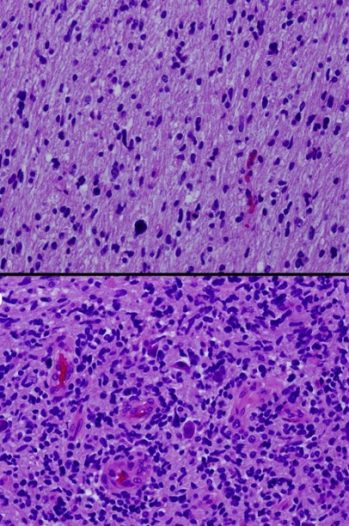
The grade of the tumor describes the level of differentiation or how “normal” the cells look when visualized under a microscope. Differentiation is an important factor in determining how likely the tumor is to grow and spread to other areas of the body. The process begins by obtaining a tumor biopsy from a patient and preparing samples either by formalin-fixation paraffin embedding (FFPE) or freezing in liquid nitrogen. The samples are then sectioned and stained, allowing the oncologist to assess the size, shape and organization of the tumor cells under a microscope. The tumor is then graded depending on the unique histology, or cell pattern. A tumor grade typically ranges from 1 (well differentiated) to 4 (undifferentiated or anaplastic). Grade 1 tumors are well differentiated, grow slowly and are considered the least aggressive. Meanwhile, tumors with grades 3 or 4 are described as undifferentiated and the most aggressive in behavior.

Different Grading Systems for Different Cancers
A score of 1 to 4 is a general approach to grading tumors. However, there are other grading systems that are more descriptive for certain cancer types. For instance, adenocarcinomas in prostate cancer have a special tumor grading system known as Gleason’s pattern. This system describes histological patterns unique for prostate cancers. A score from 1 to 5 are assigned to a primary and secondary pattern in a given tumor sample. These scores are then added together to produce a final Gleason score that ranges from 2-10. Breast cancers also have their own unique grading system known as the Nottingham histologic score. In this system, pathologists look at 3 tumor attributes: tubule formation (how abnormal is the cell structure), nuclear pleomorphism (how variable the cells look from each other), and mitotic activity (how fast the cells are dividing). A value of 1 to 3 is then assigned to each attribute where 1 is normal and 3 is irregular. The 3 scores are then added to produce the final grade that ranges from 3 to 9.

Image 1: Morphology & Grade, ICD-O-3 Morphology Codes Source

Image 2: Hematoxylin and eosin stains from different sections of a single tumor showing low-grade (Top) and high-grade (Bottom) areas. Warren, K. E. (2012). Diffuse intrinsic pontine glioma: poised for progress. Frontiers in Oncology.
Cancer Stage is More Comprehensive than Tumor Grade
A tumor grade is important in determining the potential severity of the disease. However, a more comprehensive descriptor is needed to determine cancer progression. This is where cancer staging comes in. A cancer stage not only factors in the tumor grade, but also the tumor size, position, spread, number of tumors, cell type, and involvement of neighboring lymph nodes. This information is gathered by a wide range of clinical tests including CT scans, MRI, ultrasound as well as biopsies.
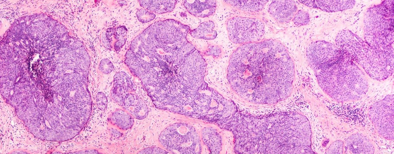
Right: Breast cancer tissue

Cancer Progression is Categorized into Four Stages
There are four stages of cancer and are depicted in roman numerals from I to IV. Stages increase as the primary tumor grows and spreads into other parts of the body. In some cases, stage 0 may be used to describe neoplastic cells that are localized and not yet cancerous. There are also more descriptive systems to stage cancer that are used by oncologists. This includes the TNM staging system (Tumor, Nodes and Metastasis) which is used for several common cancer types such as breast and colon cancer. In this system, a number value detailing the severity of the cancer is assigned to each letter. The table below describes the meaning of each value.
| Value | X | 0 | 1 | 2 | 3 | 4 |
| T (Tumor) | Main tumor cannot be measured | Main tumor cannot be found | Refers to the size/extent of main tumor. Higher the number, greater the size and spread. | |||
| N (Node) | Cancer in nearby lymph nodes cannot be measured | There is no cancer in nearby lymph nodes | Refers to the number and location of lymph nodes containing cancer. Higher the number, the more lymph nodes that contain cancer | |||
| M (Metastasis) | Metastasis cannot be measured | Cancer has not spread to other parts of the body | Cancer has spread to other parts of the body | |||
BioChain: Best-in-Class Tissue Samples
To better understand the complexities of cancer biology, scientists study a wide variety of tumor samples of different grades and cell types. BioChain offers an assortment of FFPE and frozen tissue samples that have been featured in numerous cancer publications.
In addition, tissue samples can be provided in a microarray format (TMA) with comprehensive tumor grade and TNM staging data for rapid and convenient biomarker screening (right image).
Wherever your cancer research takes you, BioChain will help you get there with their industry-leading quality research tools.
- Blog on FFPE and frozen tissue sample preparations
- Blog on tissue microarrays:

Author
Eric Torres

