Learn about DNA extraction from FFPE tissue in 10 minutes

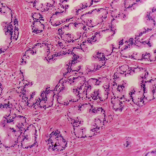
Are you a histologist looking to learn of an exciting new use for formalin fixed, paraffin embedded (FFPE) tissue blocks and sections? If so, then you are in the right place. DNA analyses such as next generation sequencing (NGS), PCR-based diagnostics, and microarray are all possible on nucleic acids extracted from this sample type.
Tissues that have been stored for an extended time are formalin fixed to keep the tissue’s protein structure intact. Then they are embedded in paraffin wax to ensure the tissue is easy to section. These FFPE tissues then can be stored for decades - creating a vast supply of samples exhibiting an array of very interesting disease states. Now, with the explosive growth of molecular genetics techniques, that require large archives of well-characterized samples, FFPE has found a new use - provided DNA of decent quality can be extracted from it. Molecular analysis of FFPE tissue promises to yield numerous new useful biomarkers for cancer and other diseases.
Until recently, nucleic acids like RNA and DNA have been difficult or impossible to isolate from FFPE tissue. Tissues prepared for more standard analysis, such as H&E staining, or IHC were not originally intended to be used in molecular analysis. Therefore, DNA (and RNA) degradation while a tissue sample sat waiting to be immersed in formalin was not on anyone's minds. Neither was it a concern that formalin tends to cross-link and degrade nucleic acids, or that embedding tissues in paraffin wax would make it difficult to extract anything out of them. Recent advances in DNA purification have made it possible for researchers to gather crucial genetic and molecular information from FFPE tissue specimens. While not discussed here, these methods for isolation of DNA can be modified for RNA, though this can be a little harder.
There are various processes used to obtain DNA from FFPE samples. Below we summarize four of most popular methods:
-
De-Waxing
The first thing that needs to happen if one is to purify DNA from FFPE tissue is to make the wax go away. It will gum up the works. Initially, toxic volatile substances, such as xylene were used to dissolve and remove the wax. Now, most protocols use non-toxic oil based agents, combined with warming the sample to remove the paraffin. BioChain's Anaprep-12 FFPE tissue DNA extraction kit uses such a non-toxic reagent.
-
Digestion with Proteinase K
Since samples will be incubated in a heat-block for the de-waxing step, the easiest way to liberate DNA from de-waxed samples is to incubate the sample with proteinase K. This will basically liquefy the tissue so that the DNA is in solution and available for purification.
-
De-crosslinking
Once a sample has been de-waxed and liquefied, the DNA will still have been crosslinked to itself and to proteins by the formalin fixation process. This crosslinking will make the DNA less pure (its stuck to protein), and also not perform well in PCR or sequencing-based applications due to the self-crosslinking issues. Fortunately, heating to 80-90 degrees C for an hour or so (depending on the protocol you're following) breaks the covalent bonds that are causing the problem. So, the first 3 steps in the DNA extraction process involve incubating the sample at elevated temperatures while occasionally adding a reagent or two. While the process takes 2-4 hours in general, the technician is free to do other things. Sort of like cooking stew in a crock pot.
-
Silica membrane based column extraction
A popular method for purifying DNA from dewaxed, digested, decrosslinked samples is to add salts and other reagents that cause DNS to bind to silica. Then the liquefied sample can be placed into a chromatography column that contains a wafer of porous silica. Either vacuum or centrifugation can be used to pull the sample through the column. Then various wash buffers can be pulled through the column to remove impurities. Lastly, water is pulled through the column in order to dissolve and recover the pure DNA. There are many commercial kits available for this purpose.
-
Magnetic silica beads
Columns can be challenging to automate, so the silica columns can be replaced by silica coated paramagnetic beads. These are basically iron beads that have been coated with silica, and will stick to a super strong magnet. Sometimes, more complex beads with a plastic core, will be coated with iron and then silica depending on the manufacturer and the purification device to be used.
The same types reagents that are used for silica column based purification will work with these magnetic beads. Instead of pulling solutions through the columns though, the prepared sample will be incubated with the magnetic beads. Then, after sufficient time for the DNA to stick to the beads a magnet will be placed next to the tube containing the solution. The beads will be pulled to the side of the tube and stay next to the magnet while the liquid containing the impurities is removed. Then the magnet can be removed from the tube, and the beads can be incubated with wash buffers. The magnet can be used again to hold the beads while the wash buffers are then removed. Finally, the thoroughly washed DNA-containing beads are incubated in elution buffer (generally water, or a very dilute Tris solution). The magnet will be used one last time to pull the beads to the side of the tube, so the purified DNA solution can be recovered.
How to increase the chances of success with FFPE samples
Various factors can affect the overall yield and quality of DNA isolated from FEPE tissues. Below are several key factors that may need to be addressed:
- Tissue specimen preparation and tissue procurement – if you can, you should fix tissues one hour after surgical resection. Then, 12-24 hours is the perfect fixation time using neutral-buffered paraformaldehyde or formalin. You should dehydrate fixed tissues thoroughly before the embedding process.
- Block storage – if possible blocks should be stored without cut faces. It prevents any damage due to exposure to water, oxygen and other factors like insects and light
- Tissue size, type, and the amount you are using for DNA isolation can have an impact. 10–20 mm is the suggested tissue thickness.
- An excess amount of paraffin – If a small tissue sample is embedded in a big paraffin block, then it's wise to trim away the extra was with a scalpel. If the tissue can't be trimmed, then increasing the amount of dewaxing agent, and/or increasing the dewaxing time may improve your results.
In conclusion, FFPE tissues that can now be used for molecular genetics analyses, and the above steps can be taken to recover nucleic acids. BioChain has a wide variety of FFPE tissues from different regions to best serve your project requirements.
Featured Products:
To learn more about the different kinds of FFPE Tissues Sections we offer, click here. If you are interested in FFPE Blocks, fill out the form below and talk to an expert!
To learn about our Automated AnaPrep System click here
https://www.youtube.com/watch?v=AMLB7fNiB7g
More information on FFPE Tissue DNA Extraction Kit


