Isolation of Mitochondria from Cells and Tissues

As mitochondria are key to cellular health, it shouldn't be surprising that an increasing number of human diseases are being traced back to these organelles. This fact leads to an intensifying need for an effective method to isolate intact mitochondria from tissues and cultured cells.
Introduction to Mitochondria:
The mitochondrion's primary function is to provide energy to eukaryotic cells. The electron transport chain, which is a form of oxidative metabolism that provides adenosine triphosphate (ATP) to power the cell, is a series of reactions that take place across the inner mitochondrial membrane. Mitochondria are present in nearly all eukaryotic cells, with the exception of some that are highly specialized, such as red blood cells. There are a few single celled fungi and protists that seem to do without mitochondria, or at least have modified versions of these organelles that do not seem to generate ATP. An example of protists that lack fully functioning mitochondria are the diplomonads, of which Giardia is an example. These organisms rely on fermentative metabolism, which does not require oxygen. There are structures that appear, visually and genetically, to have been derived from mitochondria at one point. The single celled anaerobic fungus, Microsporidium possesses an organelle called a mitosome that also appears to be derived from mitochondria. The mitosome is does not participate in oxidative metabolism, which appears to unnecessary to these obligate parasites.
History of experimenting the Isolation of Mitochondria:
Biologists have been studying mitochondria since the 1840s, when they began to be featured in drawings of high resolution microscopic images of cells. By the early 1900s, studies of extracts of guinea pig liver and minced muscle samples showed the presence of enzymes that used oxygen to produce ATP (cellular respiration), which was postulated to be a primary source of metabolic energy. By the 1940s, it was possible to fractionate cells and begin experiments that eventually determined that cell respiration occurs in the mitochondria. Currently, An increasing number of human diseases are being attributed to mitochondrial dysfunction. A list of more than 40 of these can be found on the United Mitochondrial Disease website. Although each of these diseases is relative rare, they are often fatal - which provides strong motivation to study them.
Isolation of Mitochondria from Cells and Tissues:
Mitochondrial isolation protocols involve two processes - cell disruption to break open the cells and release the cellular structures, and differential centrifugation to recover fractions that are enriched for mitochondria.
Differential Centrifugation: Differential centrifugation is the most common method of cell fractionation and mitochondrial isolation. There are numerous protocols available in the literature that provide optimized methods of employing differential centrifugation, that separate biological structures by their sedimentation coefficient - which is determined by their density and shape. Applying different levels of centrifugal force to samples in the presence of buffered salt solutions of specific densities, will cause all structures of a specific sedimentation coefficient to settle to the bottom of a collection tube at the same time, where they can be recovered. The remaining suspension of cell fragments can be subjected to centrifugation at higher g forces, to remove a second fraction, which also contains structures with sedimentation coefficients that are similar to each other. This process is repeated until a single suspension of structures from disrupted cells is separated into multiple fractions - each containing structures with related sedimentation coefficients.
Commercially available kits contain buffered salt solutions and protocols that are so highly developed that it is relatively simple to produce highly purified mitochondria preparations. Some of these kits also provide the means to break the mitochondria open and isolate the mitochondrial proteins inside. These kits should be chosen based on the simplicity of their protocols and their ability to isolate pure mitochondria fractions that remain enzymatically active.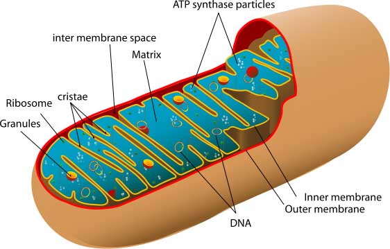
Several Tips to Isolate the Mitochondria:
The following points should be applicable to all the procedures in the isolation of mitochondria, whether one is following a protocol from a commercially available kit, or a procedure found in the literature:
- Perform all steps at 0 Degree centigrade to 4-degree centigrade.
- Work quickly and purify the mitochondria only to the degree necessary for the process. Each manipulation will result in losses.
- Keep the cell and organelle suspensions dilute during the procedure to reduce the potential for trapping and agglutination.
- Several small preparations give better yields than a single large one. Scaling up does not translate into a proportional increase in yield.
Conclusion:
Mitochondria are the source of most of the energy generated by aerobic eukaryotic organisms. There are an increasing number of mitochondrial diseases that have been identified and currently being studied - making it critical to have a good procedure for isolating intact, enzymatically active mitochondria. There published procedures for the isolation of mitochondria, as well as commercially available mitochondria isolation kits that can reduce the time required to isolate useful mitochondria fractions, and speed on one's work.
